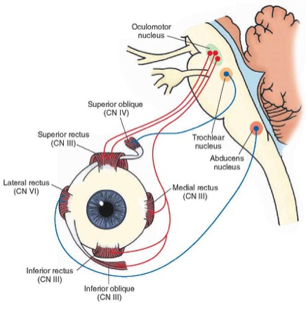
The Orbit Summary
- Identify
- Labelled
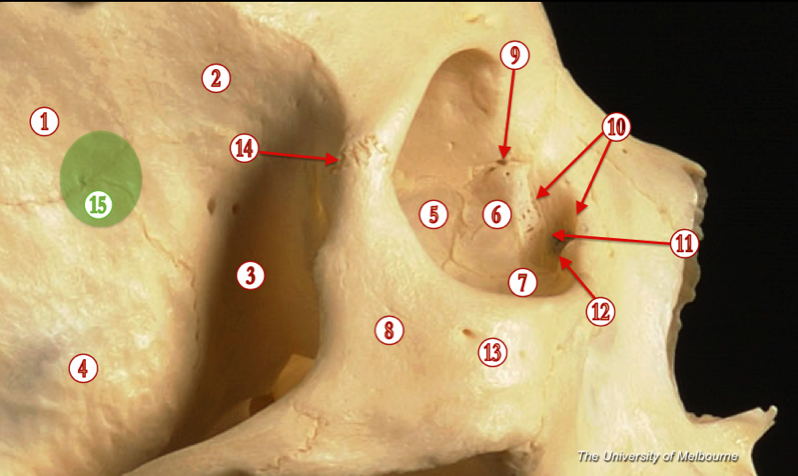
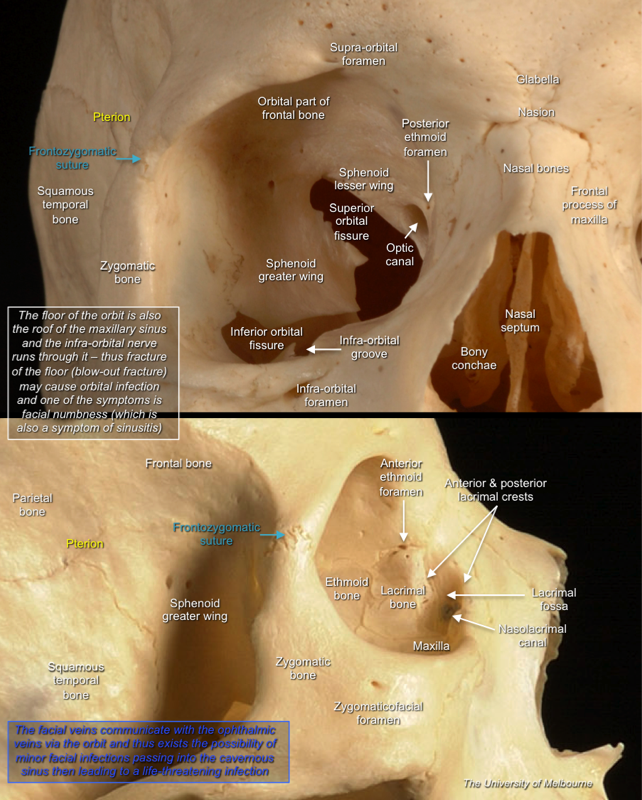
The Orbit
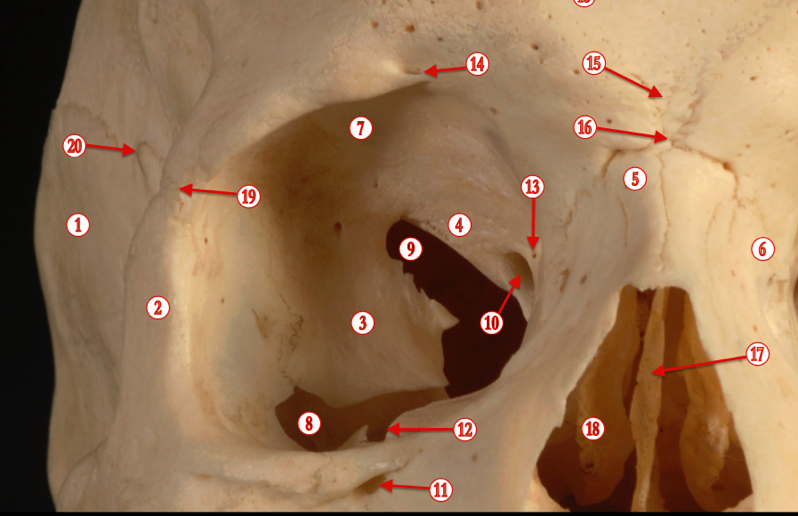
The orbits are conical or four-sided pyramidal cavities, which open into the midline of the face and point back into the head. Each consists of a base, an apex and four walls.
The apex lies near the medial end of superior orbital fissure and contains the optic canal (containing the optic nerve and ophthalmic artery), which communicates with middle cranial fossa.
The roof (superior wall) is formed primarily by the orbital plate frontal bone, and also the lesser wing of sphenoid near the apex of the orbit. The orbital surface presents medially by trochlear fovea and laterally by lacrimal fossa.
The floor (inferior wall) is formed by the orbital surface of maxilla, the orbital surface of zygomatic bone and the minute orbital process of palatine bone. Medially, near the orbital margin, is located the groove for nasolacrimal duct. Near the middle of the floor, located infraorbital groove, which leads to the infraorbital foramen. The floor is separated from the lateral wall by inferior orbital fissure, which connects the orbit to pterygopalatine and infratemporal fossa.
The medial wall is formed primarily by the orbital plate of ethmoid, as well as contributions from the frontal process of maxilla, the lacrimal bone, and a small part of the body of the sphenoid. It is the thinnest wall of the orbit, evidenced by pneumatized ethmoidal cells.
The lateral wall is formed by the frontal process of zygomatic and more posteriorly by the orbital plate of the greater wing of sphenoid. The bones meet at the zygomaticosphenoid suture. The lateral wall is the thickest wall of the orbit, important because it is the most exposed surface, highly vulnerable to blunt force trauma.
Boundaries
The base, which opens in the face, has four borders. The following bones take part in their formation:
Superior margin: frontal bone and sphenoid
Inferior margin: maxilla, palatine and zygomatic
Medial margin: ethmoid, lacrimal bone, and frontal
Lateral margin: zygomatic and sphenoid
